
Master Endodontics with Expert Guidance
Transform confusion into confidence with practical mentorship and exam preparation tailored for you.
Rated 5 stars by students
★★★★★
Empowering Dental Students with Confidence
At House of Endodontics, we transform confusion into clinical confidence through expert mentorship, practical training, and exam preparation tailored for aspiring endodontists and young dentists.
Invaluable guidance and support!
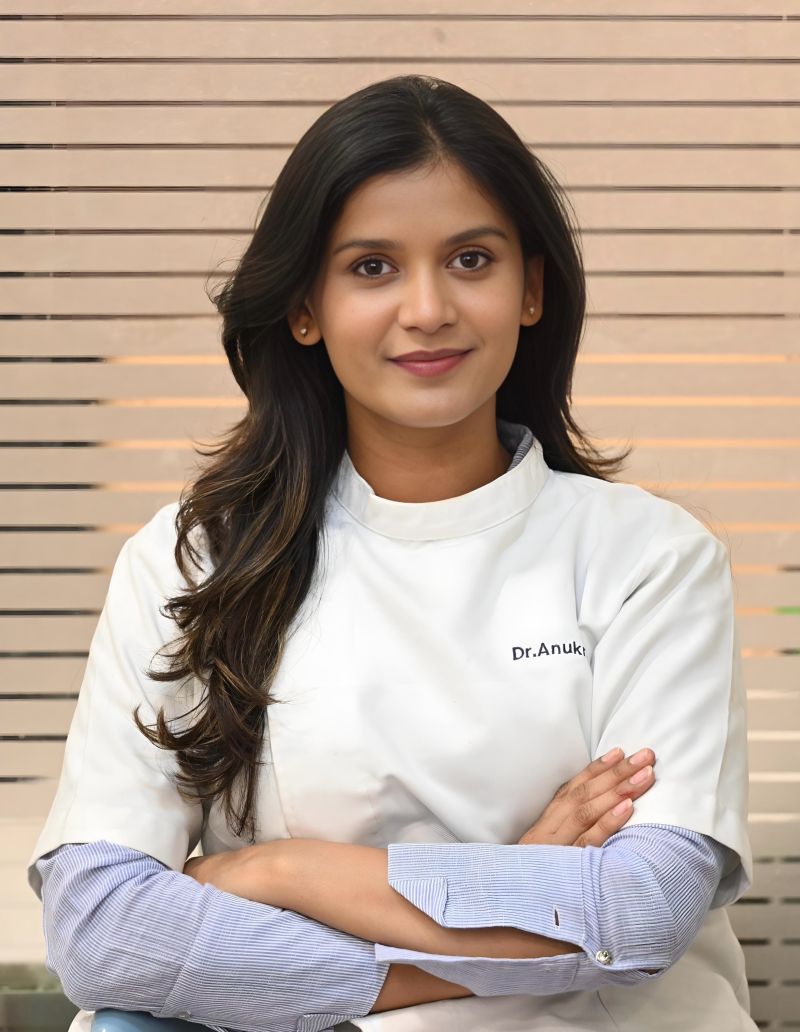
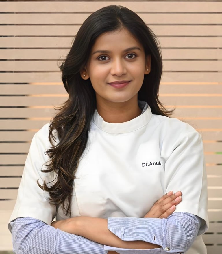
Dr. Anukrati Srivastava
"
Empower Your Practice
Transform confusion into confidence with expert mentorship and practical training in endodontics.
Mentorship Programs
Join our tailored mentorship programs designed to enhance your clinical skills and exam readiness.
Exam Preparation
Get comprehensive guidance and resources to excel in your Neet Pg exams with confidence.
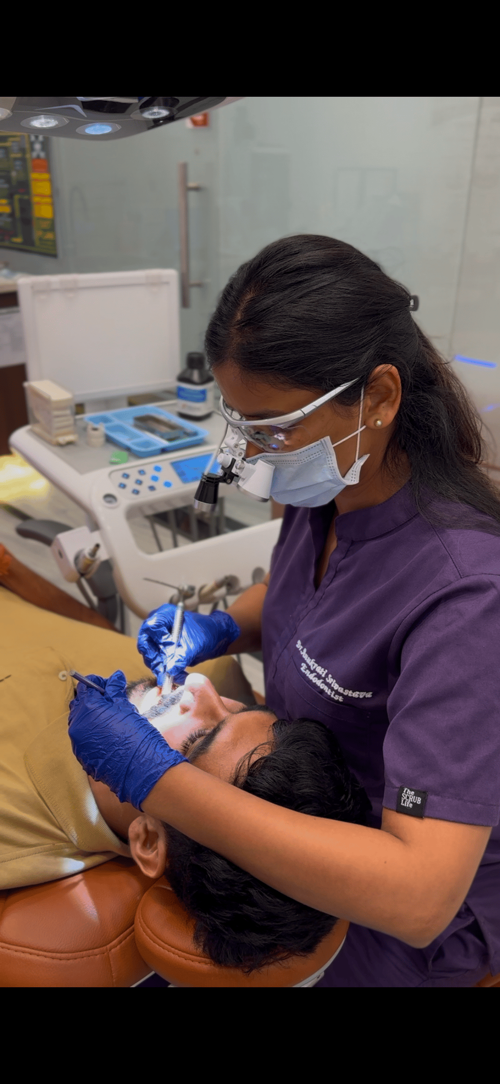
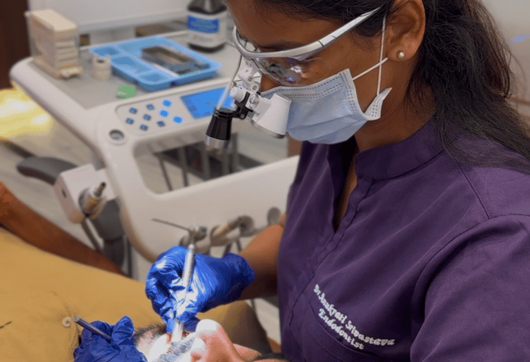
Engage in hands-on workshops that bridge theory and practice for real-world endodontic applications.
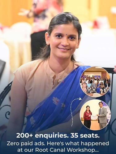
Practical Workshops

Our Workshops
Follow us on Instagram
Connect with House of Endodontics
Reach out for mentorship and guidance tailored for your endodontic journey. We're here to support your growth and confidence.
Support
+91-9587317198
+91-9481204628
houseofendodontics94@gmail.com
anukrati@houseofendodontics.com